
Arritmias auriculares
Computational Cardiology Atlas
3D Cardiac Simulator with 12-Lead ECG
Explore three-dimensional coronary anatomy, the excitation-conduction system on coronal cut, and synchronized real-time 12-lead ECG tracing with posterior (V7-V9) and right-sided (V3R-V4R) leads.
Deep Linking
Deterministic URL state architecture
Every electrophysiological scenario and ischemic pattern is encoded into unambiguous query parameters. You can share, link, or cite any clinical setup with a reproducible URL in any browser.
Reproducible example URL:
https://mioatlas.com/simulador/en/?syn=SCACEST&ter=LAD_PROX&phe=DE_WINTER&sev=SUBTOTAL
Setup: ST-elevation acute coronary syndrome, proximal left anterior descending territory, De Winter equivalent phenotype, and subtotal severity.
Valid simulator query parameters
| Parameter | Supported values | Functional description |
|---|---|---|
mode |
coronary | conduction |
Visual mode: closed coronary anatomy or coronal cut of conduction system. |
syn |
BASAL | SCACEST | SCASEST |
Clinical syndrome evaluated in coronary tree. |
ter |
LMCA | LAD_PROX | RCA_MID (18) |
Anatomical identifier of coronary stenosis or occlusion site. |
phe |
CLASSIC_STEMI | DE_WINTER | WELLENS_TYPE_A | WELLENS_TYPE_B | ASLANGER | SHARK_FIN | LBBB_SGARBOSSA | PACEMAKER_SGARBOSSA |
Electrocardiographic phenotype of acute coronary occlusion (OMI equivalents). |
ph |
HIPERAGUDA | AGUDA | ESTABLECIDA | SUBAGUDA | CRONICA |
Temporal evolutionary phase of transmural infarction. |
dom |
RIGHT | LEFT | CODOMINANT |
Anatomical coronary arterial dominance pattern. |
sev |
TOTAL | SUBTOTAL |
Severity of culprit luminal occlusion. |
add |
1 |
Enables display of additional leads (V7-V9 and V3R-V4R). |
arr |
AFIB | AVNRT | TORSADES (43) |
Identifier of rhythm disorder or conduction defect. |
43
arrhythmias with distinct morphology
18
coronary occlusion sites
8
ECG phenotypes / OMI equivalents
12+
standard leads + V7-V9 & V3R-V4R
3
coronary dominance patterns
5
temporal infarction phases
11
reconstructed coronary arteries
100%
free, no login, in-browser
Clinical Catalog
Direct access to pathological entities
Didactic selection of representative scenarios. Each button links directly to simulator state configured with exact model parameters.
Rhythm Disorders & Conduction Blocks
Arritmias auriculares
Fibrilación auricular
Arritmias de la union
Taquicardia por reentrada intranodal (lenta-rápida)
Arritmias ventriculares
Torsade de pointes
Bloqueos AV
Bloqueo AV completo (tercer grado)
Bloqueos intraventriculares
Bloqueo completo de rama izquierda
Coronary Occlusions & Myocardial Ischemia
Tronco coronario izquierdo
Tronco Coronario Izquierdo (TCI / LMCA)
Descendente anterior
Infarto anteroseptal extenso (LAD proximal, post-S1/pre-D1)
Descendente anterior
Infarto ápico-inferior (LAD recurrente / wraparound)
Circunfleja
Infarto lateral y posterolateral (LCx proximal)
Coronaria derecha
Infarto inferior aislado (RCA media)
Coronaria derecha
Infarto posterior / ínfero-septal (PDA proximal)
OMI Electrocardiographic Phenotypes
Equivalentes de oclusion (OMI)
Patrón de de Winter (Equivalente OMI)
Equivalentes de oclusion (OMI)
Síndrome de Wellens Tipo A (T Bifásica)
Equivalentes de oclusion (OMI)
Síndrome de Wellens Tipo B (T Invertida Profunda)
Equivalentes de oclusion (OMI)
Patrón de Aslanger (OMI Inferior con Enf. Multivaso)
Equivalentes de oclusion (OMI)
Criterios de Sgarbossa-Smith (Bloqueo de Rama Izquierda)
Equivalentes de oclusion (OMI)
Criterios de Sgarbossa en Ritmo de Marcapasos
Explore complete thematic catalogs
3D Catalog
Arrhythmia simulator (43 entities) →
Sinus, atrial, junctional, ventricular disorders and conduction blocks.
Ischemia Catalog
STEMI and OMI simulator →
18 coronary occlusion sites and 8 OMI equivalent phenotypes.
Structural heart conditions
Structural heart pathologies in the 3D cardiac simulator
Explore intracavitary anatomy, hemodynamics, and clinical correlation of 42 valvular, myocardial, and congenital heart diseases in an interactive 3D heart.
Technical Monitor
12-lead ECG simulator →
Continuous 120 Hz monitor, V7-V9, V3R-V4R leads, and 3D dipole.
Master Index
All 69 pathologies in one table →
Every entity with synonyms and pre-configured direct URL.
The complete MioAtlas catalog integrates 69 clinical entities modeled in three dimensions. Explanatory sheets and documentary analysis are published progressively in corresponding sections.
Perfusion & Ischemia
3D STEMI simulator & coronary ischemia
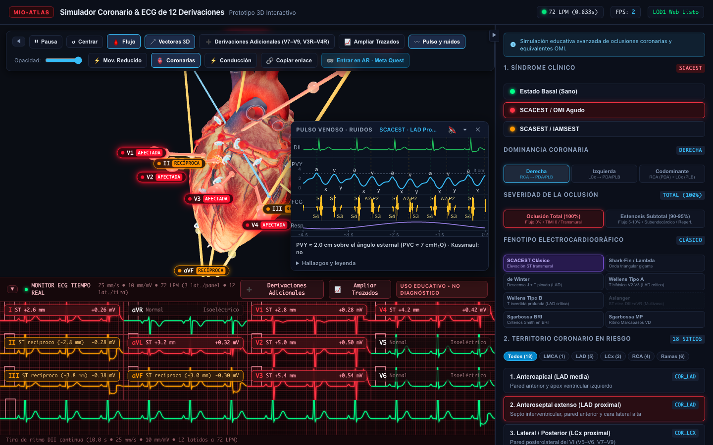
The coronary module reproduces the electrical and tissue repercussion of ST-segment elevation acute coronary syndrome (STEMI). It models 18 occlusion sites across 11 main arteries, from left main trunk to diagonal and marginal branches.
Area at risk extent and reciprocal ST elevation or depression distribution adjust dynamically according to coronary dominance anatomy (right, left, or codominant) and evolutionary phase (hyperacute, acute, fully evolved, subacute, and chronic), including critical occlusion patterns such as De Winter, Wellens, and Sgarbossa criteria.
3D Electrophysiology
Arrhythmia simulator & conduction system
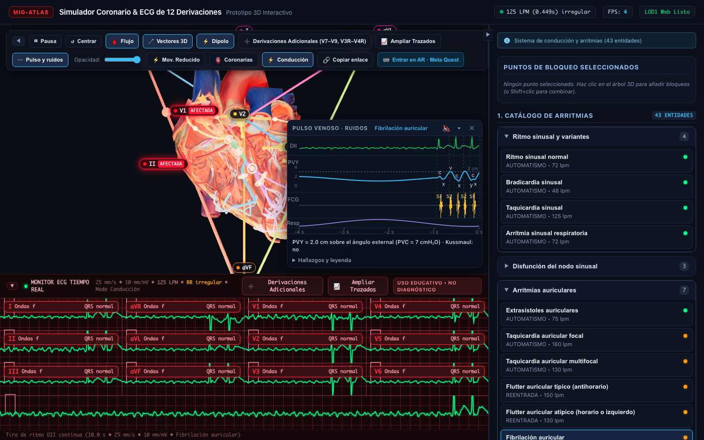
Through a coronal section of the heart, the simulator reveals the anatomical trajectory of sinoatrial node, internodal tracts, Bachmann bundle, atrioventricular node in Koch triangle, His bundle, and fascicular branches with subendocardial Purkinje network.
The electrophysiological engine reproduces 43 entities with dedicated kinematics: from physiological sinus rhythms and nodal dysfunction to atrial macro-reentries in typical flutter through cavotricuspid isthmus, chaotic activation in atrial fibrillation, AV nodal reentrant tachycardia (AVNRT), accessory pathways in Wolff-Parkinson-White, first-, second- (Mobitz I & II), and third-degree AV blocks, and bifascicular and trifascicular branch blocks.
Electrocardiographic Monitor
12-lead ECG & additional leads simulator
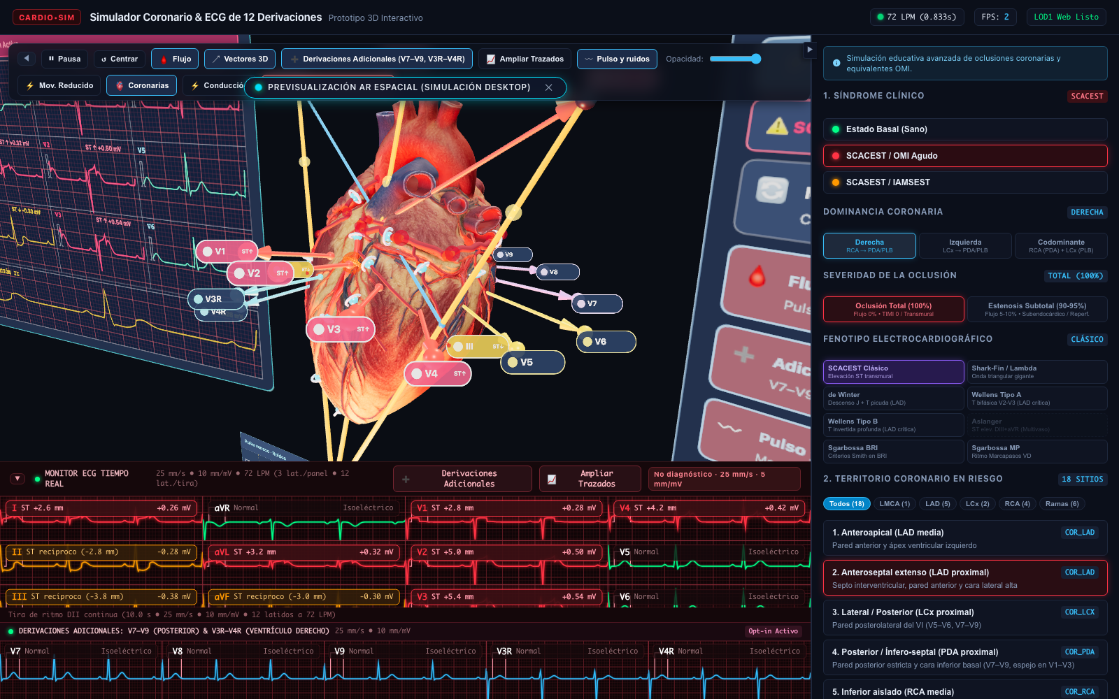
The synthetic monitor generates continuous 120 Hz tracing calibrated to standard clinical paper speed of 25 mm/s and 10 mm/mV over a millimeter grid. It projects three-dimensional lead vectors (I, II, III, aVR, aVL, aVF, and precordial V1-V6) into thoracic space with instantaneous cardiac dipole calculation.
For comprehensive evaluation of posterior wall and right ventricular infarctions, additional leads can be enabled: posterior (V7, V8, V9) and right precordial (V3R, V4R), adapting QT interval via Fridericia formulation based on instantaneous heart rate.
Jugular venous pulse & phonocardiogram
Synchronized hemodynamic correlation of the cardiac cycle. Visualization of venous pressure curve with characteristic waves (a, c, v waves and x, y descents), presence of cannon a waves in atrioventricular dissociation, and phonocardiographic recording of first (S1), second (S2), third (S3), and fourth (S4) heart sounds.
Immersive Augmented Reality (WebXR)
Native WebXR support for mixed reality devices and Meta Quest headsets. Allows projecting the heart model at 1:1 anatomical scale into physical surroundings, orbiting cavities in 3D, and examining spatial orientation of frontal and precordial lead vectors in user space.